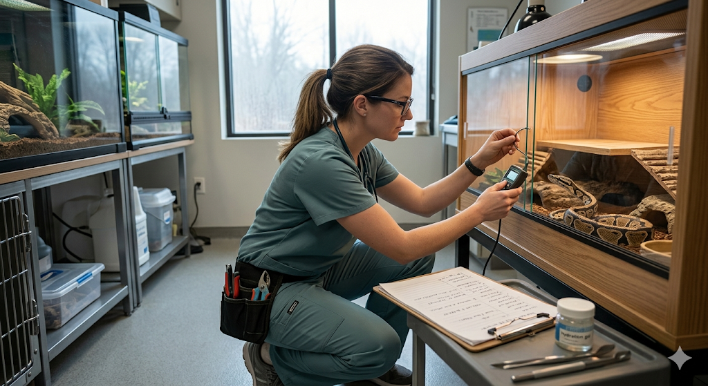
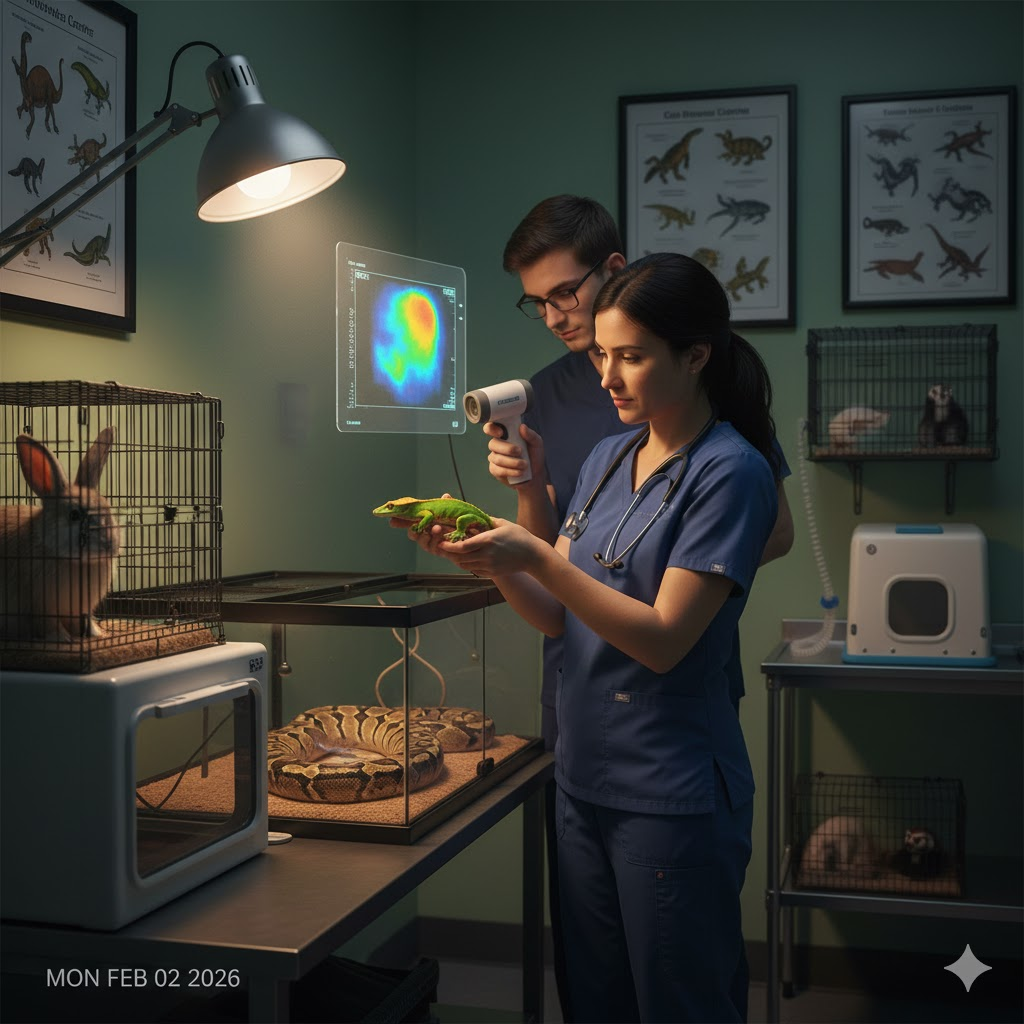
Will my pet need dental x-rays at Pet Medical Center?
You are sitting in your living room in Ames, watching your senior golden retriever try to chew a favorite tennis ball. Suddenly, he drops it, lets out a soft whimper, and paws at the side of his face. When you lift his lip, his teeth look surprisingly white, and his gums aren't even that red. You might think he just bumped his nose or is having an "off" day. However, beneath that seemingly healthy surface, a microscopic war is often being waged. In the world of 2026 veterinary medicine, we have learned that what we see with our naked eyes represents less than half of the actual story.
The most significant threats to your pet's health are often completely invisible, tucked away inside the jawbone where no amount of squinting or flashlights can reach. This is why the short answer to whether your pet needs dental x-rays at Pet Medical Center is a resounding "yes" if you want a complete picture of their well-being. Without these images, a veterinarian is essentially trying to solve a complex puzzle while wearing a blindfold.
The Iceberg Effect in Oral Health
To understand the necessity of digital dental radiographs, it helps to think of a pet's tooth like an iceberg. When you look at an iceberg from the deck of a ship, you are only seeing the small tip protruding above the waves. The massive, potentially dangerous bulk of the ice is submerged. In a dog or cat, the "crown" is the part of the tooth you can see. The "root," which is often twice as long as the crown, is submerged deep within the gingiva and the alveolar bone.
If a veterinarian only performs a visual exam, they are essentially ignoring sixty percent of the tooth's structure. Problems can fester at the root for years—causing chronic, throbbing pain—without ever showing a single blemish on the white enamel above. By the time a problem becomes visible to the eye, such as a hole in the gum or a swollen face, the damage is often catastrophic. Digital imaging acts like a high-powered window, allowing us to see through the "hull" of the jaw to inspect the foundation.
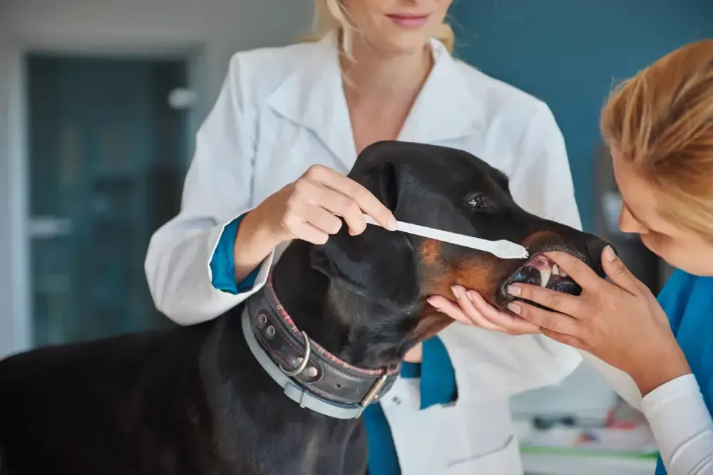
The Narrative of Hidden Issues: Why Visual Exams Aren't Enough
At Pet Medical Center in Ames, we highly recommend digital dental x-rays because they reveal hidden issues that cannot be detected during a standard physical check-up. This isn't just about finding "cavities." In fact, dogs rarely get traditional cavities like humans do. Instead, they suffer from structural failures that happen from the bottom up.
Tooth Root Abscesses
An abscess is essentially a pocket of infection that forms at the very tip of the tooth root. Think of it like a slow-burning fire in the basement of a house. The people living on the second floor might not smell smoke for a long time, but the structural beams are being weakened every minute. These infections create intense pressure within the bone. Because the jawbone cannot expand, that pressure has nowhere to go, leading to a constant, dull ache that can turn into sharp pain when the pet bites down. X-rays allow us to see the "dark halo" around a root that signals an active infection.
Bone Loss and Structural Decay
Periodontal disease is a master of stealth. Bacteria live in the "pockets" between the tooth and the gum. As they multiply, they release acidic byproducts that literally dissolve the bone. In older homes found in the Ames area, you might see wood rot that goes unnoticed until a porch step collapses. Bone loss in a pet’s mouth works the same way. We often find teeth that look perfectly stable but are actually floating in a pool of infection with no bone left to hold them in place. Imaging identifies these "periodontal pockets" and bone recession before the tooth starts wobbling.
Resorptive Lesions
This is a particularly frustrating condition, especially for our feline patients in Story County. In a resorptive lesion, the body's own cells begin to "eat" the tooth. It often starts at the root, where the tooth structure is replaced by bone-like tissue or simply disappears, leaving the sensitive nerve canal exposed to the elements. To a cat, this feels like a constant "ice cream headache" or a live wire being touched in their mouth. Without x-rays, these lesions are almost impossible to diagnose until the tooth literally fractures off at the gumline.
The 2026 Standard: Precision Planning and Better Outcomes
In the current year of 2026, the goal of Pet Medical Center is not just to "fix" problems but to plan for a lifetime of comfort. These detailed images help veterinarians diagnose and plan appropriate dental treatment before a minor issue becomes a major one.
When we have a full set of digital radiographs, we can create a "surgical map." If an extraction is necessary, the x-ray tells the doctor exactly how many roots the tooth has, whether those roots are curved like a fishhook, or if the bone around them is particularly brittle. This makes the procedure faster, safer, and much less invasive. It allows for a "measure twice, cut once" philosophy that preserves as much healthy tissue as possible.
Localization: The Ames Environment and Oral Hygiene
Living in Ames, Iowa, provides a unique context for pet ownership. Our dramatic seasonal shifts—from the humid, sweltering summers to the bone-chilling winters—affect how we interact with our pets. During the winter months, when many Ames residents are hunkered down in their homes, pets spend significantly more time indoors. This proximity often leads owners to notice "dog breath" more acutely.
However, many people attribute bad breath to the pet's diet or simply "being a dog." In reality, that odor is often the smell of volatile sulfur compounds produced by bacteria hiding under the gumline. Furthermore, Ames is home to many active pets who frequent the local parks and trails. These active lifestyles can lead to "slab fractures"—where a dog bites down on a hard rock or a frozen stick, shearing off a piece of a premolar. These fractures often look minor on the surface, but x-rays frequently reveal that the "pulp" (the tooth's internal blood and nerve supply) has been compromised, leading to a "dead" tooth that requires professional intervention.
Maintenance vs. Reactive Repair: A Comparison of Approaches
Feature
Proactive Maintenance (with X-Rays)
Reactive Repair (Visual Only)
Detection Timing
Caught in the "smoldering" stage.
Caught in the "house fire" stage.
Pain Management
Prevents pain before it starts.
Pet suffers until the issue is undeniable.
Surgical Accuracy
High; roots are mapped and understood.
Low; "blind" extractions increase risk.
Recovery Time
Usually quick; minimal tissue trauma.
Longer; often involves bone repair or multiple visits.
Long-term Value
Preserves healthy teeth for years.
Leads to premature tooth loss and systemic illness.
Systemic Protection
Stops bacteria from reaching the heart/kidneys.
Allows chronic infection to shed into the blood.
Many pet owners wonder if they can skip the imaging to save on the initial investment. While this might seem cost-effective in the short term, it almost always leads to a higher investment of time and resources later.The "Silent Sentinel": How Oral Health Protects the Body
It is a common misconception that dental issues stay in the mouth. The mouth is the most vascular part of the body, meaning it is packed with blood vessels. When your pet has an infected tooth root (which we find on those x-rays), every time they chew, they are essentially "injecting" bacteria into their own bloodstream.
These bacteria have a preference for settling on the valves of the heart and within the delicate filters of the kidneys. In 2026, we see a direct correlation between pets with neglected oral health and pets who develop early-stage kidney failure or heart murmurs. By using dental x-rays to find and eliminate these "hidden reservoirs" of infection, we aren't just cleaning teeth—we are potentially adding years to your pet's life. It is a protective shield for the entire systemic health of the animal.
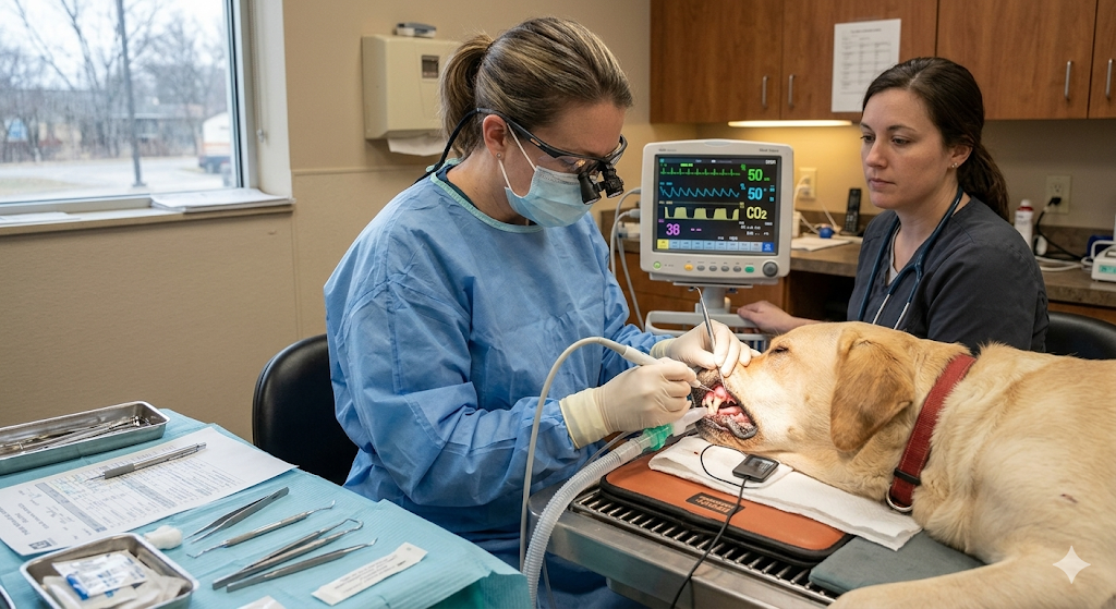
The Procedure: What Happens During Imaging?
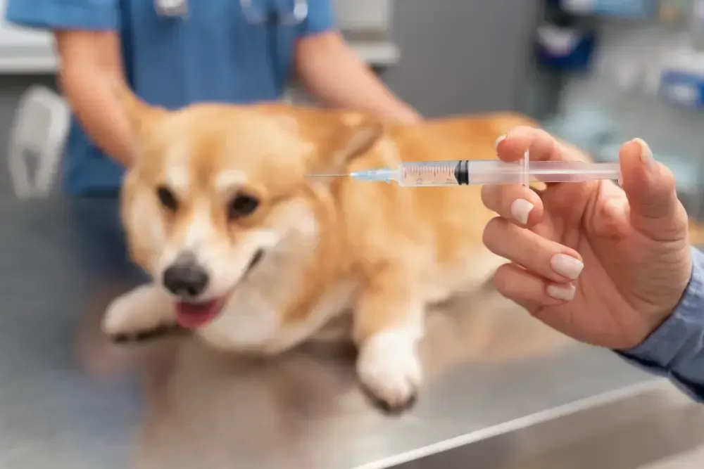
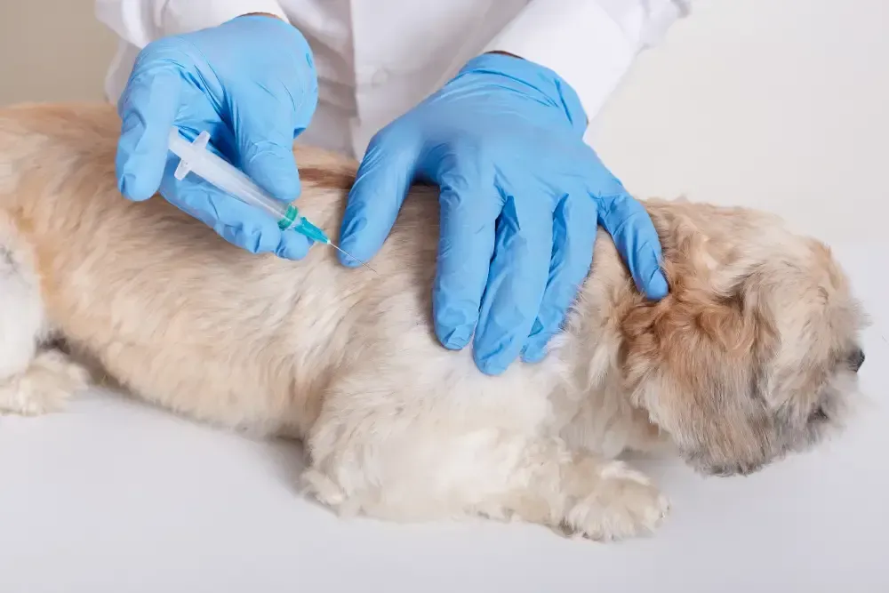
At Pet Medical Center, the process of taking dental x-rays is handled with extreme care. Because the sensor must be placed deep in the mouth and the pet must remain perfectly still to get a crisp, diagnostic image, this is performed while the pet is under a safe, monitored sedative or general anesthesia.
- Placement: A small, digital sensor (much like the ones used in human dentistry) is placed inside the mouth.
- Angle Alignment: The x-ray generator is positioned at a precise angle to avoid "foreshortening" or "elongating" the image, ensuring the root looks exactly as it does in real life.
- Instant Development: Because the system is digital, the image appears on the veterinarian's computer screen within seconds.
- Review: The doctor zooms in on the "lamina dura" (the thin line of bone surrounding the root) and the "pulp chamber" to look for any signs of widening or decay.
This process is repeated for the entire mouth. A full-mouth series is the only way to ensure nothing is missed. It is not uncommon for us to find a perfectly healthy-looking tooth on the left side and a massive abscess on the identical tooth on the right side.
The Safety of Digital Radiation in 2026
Some owners express concern about radiation exposure. However, modern digital dental x-rays use an incredibly low dose of energy. To put it in perspective, a pet would receive more radiation from a day of playing outside in the Iowa sun than they do from a full set of dental images.
The "digital" aspect is the key. Unlike old-fashioned film x-rays that required higher power and chemical processing, digital sensors are incredibly sensitive. They act like a "light sponge," soaking up the tiniest amount of energy to create a high-resolution picture. The benefit of diagnosing a hidden, painful infection infinitely outweighs the negligible risk of the imaging process itself.
Why "Anesthesia-Free" Cleanings are a Disservice
You may have seen advertisements for "anesthesia-free" dental cleanings. While these might make the teeth look white and shiny, they are purely cosmetic and, in some cases, can be harmful. It is impossible to take dental x-rays or perform a deep cleaning under the gumline on an awake pet.
Doing a cleaning without x-rays is like a dentist putting a white coat of paint over a rotted fence post. It looks better from the sidewalk, but the post is still going to snap in the next windstorm. At Pet Medical Center, we refuse to settle for "cosmetic" health. We want "structural" health, and that requires the deep-dive approach that only anesthesia and digital imaging can provide.
Understanding the "Periodontal Pocket"
One of the most frequent findings on our dental x-rays is the "periodontal pocket." When a pet has healthy gums, the tissue is tightly "shrink-wrapped" around the neck of the tooth. When bacteria move in, they act like a tiny wedge, slowly prying the gum away from the tooth.
This creates a "pocket" where food and more bacteria get trapped. Because this area is dark, warm, and lacks oxygen, the "bad" bacteria thrive. These pockets can go deep—sometimes all the way to the tip of the root—without the gumline ever looking recessed. X-rays allow us to measure the height of the bone. If the bone has "shrunk" away from the tooth, we know a pocket exists and can treat it with specialized antibiotics or deep-cleaning techniques to try and save the tooth.
The Role of Extractions: When the X-ray Says "Goodbye"
No one wants their pet to lose a tooth, but sometimes the x-ray reveals that a tooth is a "sinking ship." If a tooth has lost more than fifty percent of its bone support, or if the root is fractured, the tooth is no longer a functional tool; it is a source of chronic pain and a gateway for infection.
In these cases, the x-ray is our most valuable tool for a "clean" extraction. Some teeth, like the large "carnassial" teeth in the back of a dog's mouth, have three separate roots that spread out in different directions. Trying to pull that tooth without an x-ray is like trying to pull a stump out of the ground without knowing where the roots go. The x-ray allows the veterinarian to section the tooth into three separate pieces and remove each root individually, preventing jaw fractures and ensuring no fragments are left behind to cause future infections.
Preserving Oral Health for the Long Term
The ultimate goal of dental dentistry at Pet Medical Center is preservation. We aren't just looking for reasons to remove teeth; we are looking for ways to save them. If we catch bone loss early enough on an x-ray, we can often perform a procedure called "bone grafting" or apply a "perio-ceutic" gel that encourages the tissue to re-attach.
Without that early x-ray, we miss the window of opportunity. By the time the tooth is loose enough for a human finger to feel it, it is usually too late to save. Regular imaging is the only way to "stay ahead of the curve" and keep your pet's natural teeth in their mouth for as long as possible.
The Psychological Impact of Chronic Oral Pain
We often forget that pets are emotional beings. A dog or cat living with a hidden tooth root abscess is living in a state of "chronic stress." They may be more irritable, less likely to engage with the family, or may even develop "food aggression" because they associate mealtime with pain.
We frequently hear from Ames pet owners that their pet "acted like a puppy again" or "became much cuddlier" after a dental procedure that included x-ray-guided extractions. The pet wasn't "getting old" or "getting grumpy"; they were simply hurting. Because they couldn't tell us where it hurt, they suffered in silence. The x-ray gives them a voice. It allows them to "tell" us exactly where the pain is so we can take it away.
A Summary of the "Why" Behind the Technology
The decision to include dental x-rays in your pet's healthcare plan is a decision to prioritize their comfort and longevity. At Pet Medical Center, we believe that every pet deserves a pain-free mouth.
- Comprehensive Diagnostics: Seeing the 60% of the tooth that is hidden.
- Early Intervention: Finding "smoldering" infections before they become "house fires."
- Surgical Safety: Mapping the roots for faster, less traumatic extractions.
- Systemic Health: Preventing bacteria from traveling to the heart and kidneys.
- Quality of Life: Removing the source of chronic, hidden stress.
What Happens Next? Your Pet's Dental Journey
When you schedule a dental assessment at Pet Medical Center in Ames, we start with a thorough history and a "conscious" oral exam. We look for the "red flags" like tartar, gingivitis, or missing teeth. However, we will always discuss the "Next Step," which is the professional cleaning and imaging under anesthesia.
Once the pet is asleep and the x-rays are taken, the veterinarian will often call you to discuss the findings. We believe in a collaborative approach. We can show you the digital images on our tablets and explain exactly what we are seeing—whether it’s a healthy root or a tooth that needs to come out. This transparency ensures that you are an active participant in your pet’s health journey.
Conclusion: Investing in a Pain-Free Future
Your pet provides you with unconditional love and companionship. In return, they rely on you to be their advocate in a world they don't fully understand. They can't tell you that their left molar has a root abscess, but they can show you through their behavior and their overall health.
By choosing to include dental x-rays as part of their routine care at Pet Medical Center, you are fulfilling that role as an advocate. You are ensuring that "hidden" doesn't mean "ignored." Whether you live in a historic home near downtown Ames or a new development on the outskirts of town, your pet’s need for a healthy mouth remains the same.
Let’s move past the "visual only" era of pet care. Let’s embrace the precision, safety, and kindness of 2026 veterinary medicine. Your pet’s heart, kidneys, and—most importantly—their daily comfort will thank you for it.